Parietal Lobe Epilepsy? A Review of the Semiology of Epilepsy Surgery Cases Involving the Parietal Lobe at Westmead Hospital Between 1993 and 2018
Abstract number :
1.215
Submission category :
4. Clinical Epilepsy / 4B. Clinical Diagnosis
Year :
2018
Submission ID :
501154
Source :
www.aesnet.org
Presentation date :
12/1/2018 6:00:00 PM
Published date :
Nov 5, 2018, 18:00 PM
Authors :
Tania Farrar, Westmead Hospital; Chong Wong, Westmead Hospital; Melissa Bartley, Westmead Hospital; Mark Dexter, Westmead Hospital; Manori Wijayath, Westmead Hospital; Zebunnessa Rahman, Westmead Hospital; and Andrew Bleasel, Westmead Hospital
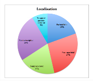
Rationale: Discrete parietal lobe epilepsy (PLE) cases are uncommon as most cases involve other cortical regions: frontal, temporal and occipital. In a large series (Salanova et al., 1995) PLE represented only 6% of patients undergoing epilepsy surgery. This paucity of cases has made a detailed assessment of parietal lobe semiology difficult. The parietal lobe contains highly eloquent areas yet the semiology of PLE is inconsistent and most often explained by the spread of the ictal activity to symptomatogenic zones beyond the parietal lobe. We review the semiology of our patients with PLE and parietal lobe epilepsy plus (PLE+) combined with their neurophysiological and neuroimaging studies to assess if common semiology patterns can be used to localize seizure onset. Methods: All epilepsy surgery cases between 1993 and 2018 were reviewed. This included stereotactic electroencephalogram (SEEG) and subdural grid (SDG) monitoring cases not proceeding to resection. There were a total of 475 cases, 268 of which were temporal lobe epilepsy (TLE) cases. Of the remaining 207 cases 87 involved the parietal lobe. These were divided into pure PLE and PLE+: temporoparietal (TPE), parieto-occipital (POE), frontoparietal (FPE) and temporo-parieto-occipital (TPOE) epilepsy, based upon intracranial EEG and surgical outcome.Clinical seizures, described by the semiological classification (Luders et al., 1998), were reviewed in detail with video EEG and intracranial EEG. MRI scans were co-registered with PET and SPECT scans, when available, and used to confirm the location of structural pathology and propagation patterns. Results: Of the 87 patients studied only sixteen (19%) had pure PLE. The most common localisations were FPE (27%), followed by POE (26%) then PLE and TPE (19% each). TPOE cases were the least common (9%).Eleven patients with pure PLE had auras, 9 were sensory and 2 were altered perception. Sensory auras although most common in PLE were not seen in all cases and were seen in all other PLE+ groups. Altered perception was seen in TPE cases. The aura types not seen in pure PLE: olfactory, cephalic and auditory auras were rare in PLE+ but visual auras were common in the PO and TPO patients. Autonomic auras were only seen in FPE cases.Seven of the 16 PLE patients had focal tonic seizures and 2 had hemiclonic seizures. The focal tonic seizures always progressed to BATS as did 1 sensory aura and 1 automotor seizure. Focal tonic seizures were most common in the pure PLE cases but did occur in TPE, POE and FPE as well, but, in the latter 2 groups, focal clonic seizures were more frequent. BATS occurred in FPE, PLE and POE groups more often than in the TPE group and were not seen at all in the TPOE.In the PLE group, 1 patient had a dialeptic seizure and 4 had automotor seizures; 4 of these progressed to versive seizures. Dialeptic seizures were rare and seen in only 3 of the 71 patients with PLE+. Automotor seizures, in contrast were seen commonly in all groups but more so in PLE+ cases than in pure PLE patients. Versive seizures occurred in all groups but were most common in the POE and FPE. Hypermotor seizures were rare and seen in only 1 of the PLE patients and 3 of the FPE patients. Conclusions: There does not appear to a distinct semiology in PLE. Like researchers before us we found that the semiology of the seizures was extremely diverse and the majority of symptoms were due to the spread of ictal activity outside of the parietal lobe, most commonly the temporolimbic or mesial frontal regions. While sensory auras and tonic seizures were the most common features seen in the PLE group they were not seen in all patients and were seen in the other groups studied. Funding: Nil

.tmb-.png?Culture=en&sfvrsn=3ccae870_0)