CSF Suppressed T2-Weighted Imaging at 7T in Epilepsy
Abstract number :
2.178
Submission category :
5. Neuro Imaging / 5A. Structural Imaging
Year :
2018
Submission ID :
502143
Source :
www.aesnet.org
Presentation date :
12/2/2018 4:04:48 PM
Published date :
Nov 5, 2018, 18:00 PM
Authors :
Chan H. Moon, University of Pittsburgh; Arun Antony, University of Pittsburgh; Alexandra Urban, University of Pittsburgh; Joanna Fong, University of Pittsburgh; Naoir Zaher, University of Pittsburgh; R. Mark Richardson, University of Pittsburgh; Anto Bagi
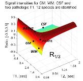
Rationale: 7T imaging has been used to acquire high resolution T1 weighted imaging, which can be helpful to epilepsy neuroimaging. However, because of the high RF power requirements for 7T imaging, FLAIR contrast is commonly acquired at lower resolution (particularly for slice thickness) and partial brain coverage. A novel imaging strategy (magnetization prepared fluid attenuated gradient echo, MPFLAGRE) to generate high resolution CSF suppressed T2-weighted contrast at 7T is described and shown in controls and localization related epilepsy patients. Methods: The MPFLAGRE methodology uses a driven equilibrium spin echo preparation within an inversion recovery with multiple gradient echo blocks. Images are combined via the self-normalization approach, which achieves CSF suppression through proper timing of individual blocks and minimizes sources of variation. Simulation and in vivo demonstrations in epilepsy patients are shown with measurement of contrast-to-noise ratios in n=5 healthy controls. We used a Siemens whole body 7T Magnetom 8 channel multiple transmit system with body gradient coil and an 8x2 transceiver array for all acquisitions. With the performance of the transceiver, conventional pulses modules are used giving a global SAR of less than 75% of FDA guidelines. A GRAPPA factor of 3 was used, achieving a 0.7 x 0.7 x 1.2mm (<0.6mm3) resolution with an acquisition time of <10min. Results: Fig. 1 shows the T2 and T1 dependence of the MPFLAGRE contrast, and shows the steep dependence of signal intensity over pathologic T2 values. From n=5 healthy controls, thalamic and white matter CNR (taken in ratio to CSF) was 6.6±1.0 and 6.9±1.0 respectively, the small difference between white and gray matter reflecting the known small T2 differences between WM, GM at 7T. Fig. 2 shows the MPFLAGRE-4 images from two epilepsy patients. Overall, the data shows that there is enhanced signal intensity at the cortical rim and ependymal, consistent with the report of von Veluw 2014 who concluded that increased T2 contributes to their enhanced FLAIR signal intensity. While quantitative T2 data do not exist on these tissue components, a long T2 for the cortical rim is not surprising given that it contains the comparatively poorly vascularized molecular layer I of the neocortex. Similarly, the high signal intensity from the ependyma is likely related to its function as the neuroepithelial layer around the ventricle that contributes to the production and regulation of CSF. In Fig. 2 (left A-C), the patient has known neocortical epilepsy most likely temporal lobe and shows bright signal in the lateral temporal neocortex and hippocampus, this is not well identified in the clinical 3T FLAIR. The right patient data (D) had had a seizure within 8 hours of imaging, and showed a widespread increased MPFLAGRE intensity through much of the left temporal lobe. The conspicuity of detection by the MPFLAGRE sequence is likely related to the steep dependence of the signal between normal to pathologic T2s as shown in the simulation (FIg. 1). Conclusions: FLAIR imaging generally plays an important clinical role for lesion detection based on pathologic T2 signal increase. At 3T, FLAIR images are commonly acquired with comparatively lower resolution (particularly in slice thickness) because of the needed lengthy acquisition times, this problem being exacerbated at 7T due to power limitations. We have developed and demonstrated the MPFLAGRE sequence as a flexible acquisition to generate T2 and CSF suppressed T2-weighted contrast at 7T. This first experience of the high resolution 7T MPFLAGRE sequence in epilepsy patients suggests that it may be useful in identifying regions involved in the seizure. Funding: This work supported by NIH R01EB011639, R01NS090417 and R01NS081772.

.tmb-.jpg?Culture=en&sfvrsn=7ee41e68_0)