Frontal Lobe Seizure Differentiated From Non-Epileptic Seizure by 256 Channel dEEG
Abstract number :
2.040
Submission category :
3. Neurophysiology / 3C. Other Clinical EEG
Year :
2018
Submission ID :
501591
Source :
www.aesnet.org
Presentation date :
12/2/2018 4:04:48 PM
Published date :
Nov 5, 2018, 18:00 PM
Authors :
Hisanori Hasegawa, University of South Carolina School of Medicine in Greenville and Chaim Colen, Beaumont Hospital in Gross Point
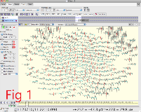
Rationale: Frontal lobe epilepsy is occasionally difficult to distinguish from non-epileptic seizures. It is not uncommon for a patient who has frequent seizure attacks to experience correlating epileptiform discharges during ictal events in CCTV-EEG. Some patients may be diagnosed as a non-epileptic seizure just because of the absence of EEG changes. Diagnosis may remain inconclusive and treatment may not be decisive in that situation. Conventional 10-20 system EEG has a high rate of false negative in detection of the frontal seizure focus. dEEG (dense array EEG) could detect seizure focus better by higher spatial resolution if it may be available in selected patients. Methods: This is a retrospective study. The patients included in this study are cases who were admitted to EMU for intractable seizure episodes in phase I, and had negative diagnosis of non-epileptic seizure. These patients were advised to undergo a 60 min recording of dEEG study. The studies were performed by 256 channel EGI System (Eugene, OR). Inter-electrode distance in dEEG is 2 cm in average while that of 10/20 System is 6.5 cm. Results: Eight patients who failed to demonstrate seizure findings in scalp recordings had dEEG evaluation.Patient 1 presented with semiology of mesial frontal lobe seizure. CCTV-EEG was non-epileptic. dEEG demonstrated low amplitude sharp interictal discharges in left inferior frontal leads. (fig 1). Depth electrodes confirmed mesial frontal seizure.Patient 2 presented with semiology of lateral frontal neocortical seizure. CCTV-EEG was normal. dEEG demonstrated right frontal electrographic seizure. (fig 2)Patient 3 presented with semiology of anterior frontal epilepsy. dEEG demonstrated right inferior frontal focus with DC shift at onset.Patient 4 presented with semiology of orbitofrontal epilepsy. dEEG demonstrated left inferior frontal focus.Patient 5 presented with semiology of insular epilepsy. dEEG demonstrated right anterior temporal localization.Patient 6 presented with semiology of frontal epilepsy. dEEG was normal.Patient 7 presented with semiology of frontal epilepsy. dEEG was normal.Patient 8 presented with semiology of frontal epilepsy. dEEG was normal. Conclusions: Five patients out of eight revealed abnormal focal findings in dEEG. Two of the five patients ictal events with epileptiform discharges. The study suggests that utilization of 60 minutes dEEG recording may better diagnose frontal lobe epilepsy in patients who were regarded as having PNES due to the lack of correlating EEG findings in 10/20 System. Analyzing seizure semiology may be helpful to target area of localization in dEEG recording. The limitation is that mesial frontal seizure is still difficult to identify by dEEG alone. Funding: This study did not receive financial support from any third party.

.tmb-.png?Culture=en&sfvrsn=373266eb_0)