LOCATING SUBDURAL ELECTRODES IN CT IMAGES USING 3-D SURFACE RECONSTRUCTION
Abstract number :
2.325
Submission category :
Year :
2004
Submission ID :
4774
Source :
www.aesnet.org
Presentation date :
12/2/2004 12:00:00 AM
Published date :
Dec 1, 2004, 06:00 AM
Authors :
1,2John D. Hunter, 2Jake Reimer, 2Diana M. Hanan, 1Kurt E. Hecox, and 1,2,3Vernon L. Towle
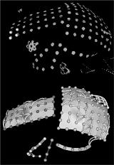
Chronically implanted subdural electrodes are increasingly being used in invasive epilepsy evaluations as a means to locate intractable epileptic foci. In difficult multifocal cases, often more than 100 electrodes are implanted. Accurately determining the location of the electrodes in 3-D space in a timely manner is a critical first step in interpreting the electrocorticographic (ECoG) findings for planning seizure focus resections. Accurate localization and identification has traditionally been ignored, or at best is a tedious process, taking a trained technologist up to five hours per patient. Previously, we developed an interactive application in which the electrodes could be marked in image plane slices from the CT. We present an extension of this method in which surface rendering techniques are used to reconstruct the electrode surfaces automatically from high resolution CT scans (1 mm slices). After identifying the intensity of the electrodes in the scan, the rendering algorithm parameters are set to eliminate everything but the electrodes from the rendered image. The resulting objects are then filtered and segmented to eliminate other metallic artifacts and viewed in a 3-D window. A ray casting technique is used to define the spatial coordinates of the rendered electrodes interactively. The application runs across operating systems platforms and is developed solely with open source tools free for non-commercial use. The new procedure produces accurate results (less than half the width of an electrode) validated against visual localization and intraoperative photographs in a small fraction of the time. The localization of electrodes allows for the control of distance effects on coherence. Using this information, we present findings from an analysis of the spatial distribution of coherence and spectral power in 15 surgery candidates with intractable epilepsy.[figure1] This technique dramatically reduces the time required to accurately identify subdural electrodes locations, increasing their value for both clinical evaluations and research applications, such as source localization. (Supported by NIH NS40514-02)
