Reevaluation of Postoperative Long-Term Epilepsy-Associated Brain Tumors
Abstract number :
1.362
Submission category :
9. Surgery / 9C. All Ages
Year :
2018
Submission ID :
501388
Source :
www.aesnet.org
Presentation date :
12/1/2018 6:00:00 PM
Published date :
Nov 5, 2018, 18:00 PM
Authors :
Wenbo Zhang, Minnesota Epilepsy Group, P.A.; Jason Doescher, Minnesota Epilepsy Group, P.A.; Nitin Agarwal, Minnesota Epilepsy Group, P.A.; Michael Frost, Minnesota Epilepsy Group, P.A.; and Deanna Dickens, Minnesota Epilepsy Group, P.A.
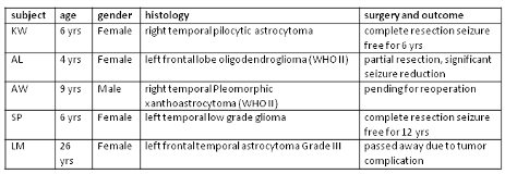
Rationale: About 60% of brain tumor patients will experience seizure at least once during their illness. Primary brain tumors account for 5% of new onset seizures in adults and over 10% of lesional focal epilepsies. The goal to treat these patients includes: improve survival rate, seizure control and quality of life. This can potentially be achieved with total tumor resection. We reviewed 5 patients who had resective surgery of the low grade glial or neuroglial tumor, later they were re-evaluated for epilepsy surgery due to the medically refractory seizures. Methods: We reviewed 5 patients who had resective surgery of the low grade glial or neuroglial tumor, later they were re-evaluated for epilepsy surgery due to the medically refractory seizures (see Table). There are 4 female and one male patients, age of 4, 6, 6, 9 and 26 years. The reevaluation included structural MRI with epilepsy protocol, long term video EEG recording, MEG as well as invasive ECoG (2 patients). Results: Three patients underwent further resective surgery, 2 patients have been seizure free after the complete resection of epileptogenic region defined by structural MRI, MEG and invasive EEG; one patient experienced partial resection to avoid neurological deficit, her seizure has been improved significantly; one patient with 2 previously “gross total” resection, is pending for another surgical intervention. One patient could not precede for resective surgery due to potential neurological deficit.Figure. Pt KW. Top panel – MR images after first tumor resection surgery of right temporal lobe; middle panel - MSI interictal spikes at posterior resection margin; lower panel – extended right temporal lobectomy plus hippocampus resection demonstrated on MR images after the 2nd surgery. Conclusions: Conclusion: Based on our limited data, we recommend that epilepsy-associated brain tumor should be evaluated with both structural imaging and EEG/MEG. If possible, complete and generous resection is performed to improve seizure control and quality of life. Funding: Not applicable

.tmb-.png?Culture=en&sfvrsn=d7189bad_0)