THE EFFECTS OF LAMOTRIGINE AND TOPIRAMATE ON CEREBRAL GLUCOSE METABOLISM
Abstract number :
2.293
Submission category :
Year :
2004
Submission ID :
782
Source :
www.aesnet.org
Presentation date :
12/2/2004 12:00:00 AM
Published date :
Dec 1, 2004, 06:00 AM
Authors :
1Eun Yeon Joo, 1Woo Suk Tae, 1Jee Hyun Kim, 1Sujung Choi, 2Byung Tae Kim, and 1Seung Bong Hong
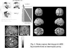
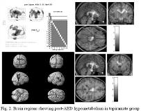
Antiepilpetic drugs (AED) are known to have inhibitory effects on brain. To investigate the effects of lamotrigine and topiramate on cerebral metabolism, we performed 18F-fluorodeoxy glucose positron emission tomography (FDG-PET) in patients with new-onset epilepsy. FDG-PET were performed two times (before and after AED medication) in 13 patients (Topiramate group, M/F=9/4, 28.2[plusmn]11.4 years) and 11 patients (Lamotrigine group, M/F=5/6, 29.1[plusmn]10.4 years). For SPM analysis, all PET images were spatially normalized to the standard PET template, then smoothed with 12-mm full width at half maximum gaussian kernel. The [italic]paired t[/italic]-test was performed for comparison between pre- and post-AED PET images. The height threshold was set to false discoveryrate (FDR) corrected [italic]P[/italic][lt]0.05, and extent threshold was set to [italic]K[/italic][sub]E[/sub] [gt]100. SPM analysis between post- and pre-AED FDG-PETs in lamotrigine group showed hypometabolism in both inferior temporal gyri, left superior frontal gyrus, both superior parietal lobules, right hippocampus, left para hippocampal gyrus, left lingual gyrus, both putamens, both caudate nuclei, left thalamus, both hypothalamus, left midbrain (corrected [italic]p[/italic][lt]0.05)(Fig. 1) whereas that in topiramate group showed hypometabolism in both parietal lobules, left inferior posterior temporal gyrus, both superior anterior frontal gyri, left middle frontal gyrus, both midbrains, both caudate nuclei, both thalami, right cingulate gyrus, corpus callosum, and the white matters of both parietal lobes and right temporal lobe (corrected [italic]p[/italic][lt]0.05)(Fig. 2). There was no brain regions showing post-AED hypermetabolism.[figure1][figure2] Lamotrigine decreased glucose metabolism more in cerebral cortex (both inferior temporal and parietal lobes), less in deep gray and white matters while topiramte decreased glucose metablism more in corpus callosum, thalamus, white matters and midbrain, less in cerebral cortex. (Supported by The NRL grant 2000-N-NC-01-C-163(2003) by the Korean Ministry of Science and Technology, and the grant A18-01-00 from the next-generation new technology development program of the Korean Ministry of Commerce, Industry and Energy.)

