VOXEL-BASED OPTIMIZED MORPHOMETRY (VBM) IN GRAY AND WHITE MATTER IN TEMPORAL LOBE EPILEPSY (TLE)
Abstract number :
1.089
Submission category :
Year :
2005
Submission ID :
5141
Source :
www.aesnet.org
Presentation date :
12/3/2005 12:00:00 AM
Published date :
Dec 2, 2005, 06:00 AM
Authors :
1Susanne G. Mueller, 2Kenneth D. Laxer, 1Nate Cashdollar, 1Derek L. Flenniken, and 1Michael W. Weiner
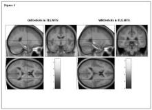
In TLE with evidence for hippocampal sclerosis (TLE-MTS) volumetric gray (GM) and white (WM) matter abnormalities are not restricted to the hippocampus but are also found in extrahippocampal structures involved in seizure spread. Less is known about extrahippocampal volumetric abnormalities in TLE without hippocampal sclerosis (TLE-no). In this study we used optimized VBM with and without modulation with the following aims: 1. To confirm extrahippocampal GM and WM abnormalities found in TLE-MTS by previous VBM studies. 2. To seek evidence of extrahippocampal GM and WM abnormalities in TLE-no. 3. To compare extrahippcampal GM and WM abnormalities in TLE-MTS and TLE-no. Optimized VBM of gray (GM) and white matter (WM) with and without modulation was performed in 26 TLE-MTS (mean age 35.6[plusmn] 9.7) and 17 TLE-no (mean age 35.6[plusmn] 11.1) and 30 healthy controls (mean age 30.3[plusmn] 11.1) using the SPM2 software (Wellcome Department of Cognitive Neurology). A symmetrical custom template and symmetrical gray, white and CSF priors were created and images of left TLE patients were side flipped so that the focus was on same side in all patients. All images were smoothed by a 4 mm FWHM kernel. Results were corrected for multiple comparisons at FDR p[lt]0.05. TLE-MTS: GM volume reductions: Ipsilateral hippocampus, fusiform gyrus, posterior cingulate, bilateral medial and posterior thalamus, bilateral cerebellum and parieto-occipital GM. GM concentration reductions: Ipsilateral hippocampus, fusiform gyrus, bilateral cerebellum. WM volume reductions: Ipsilateral parahippocampal gyrus, temporal lobe and temporal stem, posterior internal capsula, bilaterally in cerebellum, splenium and pons/midbrain. WM concentration reduction: Similar locations as WM volume reductions, less extensive. TLE-no: No GM concentration/volume reductions or WM concentration/volume reductions compared to controls. There were also no differences in GM/WM volumes/concentrations between TLE-MTS and TLE-no. (Figure 1) In TLE-MTS VBM showed extensive GM and WM volume reductions in the ipsilateral hippocampus and in extrahippocampal regions connected to the mesio-temporal lobe and involved in seizure spread. The lack of hippocampal GM/WM deficits in TLE-no had to be expected but in addition to this, this group has also no or at least no uniform GM/WM deficits in brain regions connected to the presumed epileptogenic focus. This further supports the hypothesis that TLE-no is an entity distinct from TLE-MTS.[figure1] (Supported by RO1-NS31966 to KDL.)
