EEG and Cardiac Correlates in Drug-Resistant Temporal Lobe Epilepsy
Abstract number :
3.054
Submission category :
1. Basic Mechanisms / 1D. Mechanisms of Therapeutic Interventions
Year :
2018
Submission ID :
506878
Source :
www.aesnet.org
Presentation date :
12/3/2018 1:55:12 PM
Published date :
Nov 5, 2018, 18:00 PM
Authors :
Eline Mesquita Melo, University Federal of Para; Rodrigo Pereira, Institute of Technology, Federal University of Pará, Belém (PA); José Fiel, Institute of Technology, Federal University of Pará, Belém (PA); Raphael Navegantes, Ins
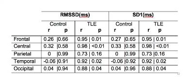
Rationale: Some neurological disorders are associated with dysfunction of cardiac autonomic control. For instance, heart rate variability (HRV), measured by the RMSSD (root-mean square differences of successive R-R intervals) and SD1, a poincaré plot component, is reduced in patients with refractory Temporal Lobe Epilepsy (TLE). Such patients also display abnormalities in the electroencephalographic (EEG) alpha rhythm (8-13 Hz), including a reduction in its power peak. The alpha rhythm plays a role in cognitive, psychomotor, psycho-emotional aspects of human life and its associated abnormalities in epileptic patients is of therapeutic interest. Methods: Nine minutes of resting state EEG was recorded from 5 patients (aged 33±12 years) with a diagnostic of drug-resistant temporal lobe epilepsy (TLE). A control group comprised of 5 age-matched healthy subjects was submitted to the same procedure. EEG was recorded with a 22-channel system (Neuromap 40i, Neurotec) with a sampling frequency of 256 Hz and using Fpz as ground. Channels were referenced to the linked mastoids. The recordings were pre-processed to remove biological artifacts. The mean power of the lower-alpha band was computed using the EEGLAB matlab toolbox. The EKG signals were analyzed with the Kubius HRV software (3.0.1/Kubos). We measured the correlation between the HRV parameters RMSSD and SD1 and the mean EEG lower-alpha power in the following regions of interest (ROI) (with corresponding electrodes): Frontal (Fp1, Fp2, F3, F4, F7, F8, Fz), Central (C3, C4, Cz), Temporal (T3, T4, T5, T6), Parietal (P3, P4, Pz) and Occipital (O1, O2, Oz). Results: The correlation between lower-alpha power and HRV was statistically significant only for TLE subjects and in the following ROIs: Frontal, Central, Temporal and Occipital (see table 1). Conclusions: Our results show that individuals with TLE display a shift in resting lower-alpha power towards lower values. This may be the result of chronic use of epileptic medication. Our data confirm previous findings in the literature showing that HRV has a tendency towards lower values when compared with controls. Finally, we show a strong correlation between HRV and mean lower-alpha power in TLE subjects. This latter finding suggests that autonomic and cortical function is tightly linked in TLE patients. The consequences of this phenomenon are still unknown and should be further investigated. Funding: No financial support
