ICTAL AND POSTICTAL DYSPROSODIAS: CLINICAL FEATURES AND DOCUMENTATION BY ACOUSTIC ANALYSIS
Abstract number :
2.204
Submission category :
Year :
2004
Submission ID :
4726
Source :
www.aesnet.org
Presentation date :
12/2/2004 12:00:00 AM
Published date :
Dec 1, 2004, 06:00 AM
Authors :
Sebastian Gürtler, and Alois Ebner
Ictal and postictal dysprosodias are rarely reported but a phenomenon which is probably recognized too infrequently. In other neurological conditions dysprosodias are reported mainly after lesions of the right hemisphere, and so seizure-related dysprosodias may serve as a clinical lateralizing sign to the non-dominant hemisphere. Aim of this study was to collect the clinical features of patients with seizure-related dysprosodias and to characterize dysprosodic symptoms. We identified retrospectively 26 patients with a seizure-related dysprosodia who had undergone presurgical evaluation. We documented EEG seizure patterns and epilepsy syndromes and we differentiated between postictal and ictal dysprosodia. We also analyzed available video recordings of seizures and classified by listening to the recordings and using Fourier spectral analysis. In 69 % of the patients dysprosodia was found as an ictal symptom, in 31 % postictal. The syndromes were 8 % focal epilepsies without clear lateralization, 69 % right and 12% bilateral temporal lobe epilepsies, 8% right frontal lobe epilepsies and one (3%) suspected left frontal epilepsy. Seizure patterns in 92 % were on the right hemisphere, 8 % of the patients showed no EEG seizure pattern during ictal dysprosodia, including the patient with the left frontal lesion.
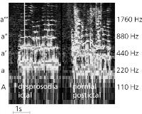
Each patient had stereotyped patterns of dysprosodia, but there was a big variability between the patients. Changes occur on four axes: 1) Pitch: We saw an exaggeration of a likely normal pitch course, a continuous increasing of pitch during one utterance or a flattened pitch course (see figure). 2) Volume: especially in ictal speech we found an increased intensity of speech, but decreased intensity of speech down to aphonia also occured. 3) Time course: We found some syllables prolonged or stops after each syllable, which could have resulted in a phase of stuttering. 4) Resonances: A few patients showed changes in the harmonic spectrum resulting in an abnormal nasal or in hollow speech.
Often the pathology of prosody was only obvious in comparison with the interictal speech.[figure1] Seizure-related dysprosodias are not uncommon and seem to occur nearly exclusively in right hemisphere seizures originating in the frontal and temporal lobe. Nevertheless, the value as a clinical lateralizing sign still has to be determined prospectively, for dysprosody may be underdiagnosed in patients with aphasic speech disturbances. Fourier analysis can especially help to measure pitch changes. Seizure-related dysprosodias may serve as a model for changes of prosody because they allow comparisons within single subjects.
