Longitudinal Microstructural Changes Following Status Epilepticus in the Rat Pilocarpine Model of Epilepsy
Abstract number :
2.185
Submission category :
5. Neuro Imaging / 5A. Structural Imaging
Year :
2018
Submission ID :
502572
Source :
www.aesnet.org
Presentation date :
12/2/2018 4:04:48 PM
Published date :
Nov 5, 2018, 18:00 PM
Authors :
Luis Concha, Universidad Nacional Autonoma de Mexico; Luis Cuauhtemoc Marquez-Bravo, Universidad Nacional Autonoma de Mexico; and Hiram Luna-Munguia, Universidad Nacional Autonoma de Mexico
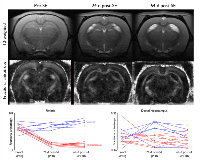
Rationale: Diffusion Tensor Imaging (DTI) provides insight into tissue microstructure and has been used to demonstrate extensive white matter abnormalities in patients with temporal lobe epilepsy (Otte et al., Epilepsia 2012;53:659-667). In the fornix, in particular, these abnormalities correspond to reduced axonal density and alterations of the myelin sheaths (Concha et al., J Neurosci 2010;30:996-1002). Nevertheless, the timing of establishment and development of these alterations is unclear. In the present study, we aimed to identify progressive white and gray matter changes of the hippocampal formation after status epilepticus (SE). Methods: SE was induced by intraperitoneal pilocarpine on male Sprague-Dawley rats (Luna-Munguía et al., Sci Rep 2017;7:1311) in order to generate epileptic (n=9) and control (n=6) animals. All animals were imaged using a 7 T scanner at three time-points: 16 days prior to SE induction, and 24 and 64 days post SE (pixel resolution=133x135 µm² per side, slice thickness = 750 µm (slice gap = 50 µm); two diffusion-weightings (b=650 and 2000 s/mm²), each with the same 40 diffusion encoding directions. Fractional anisotropy (FA) and mean diffusivity (MD) were calculated bilaterally at the fimbria/fornix (FF), and the dorsal and ventral hippocampus (DH and VH, respectively). T2 signal intensity changes (relative to CSF) were also measured in the same regions using a non-diffusion-weighted image normalized with respect to cerebrospinal fluid intensity. Results: DTI parameters and T2 signal intensity did not change over time in control animals. Contrastingly, experimental animals displayed conspicuous T2 signal hyperintensities in both hippocampi and fimbria/fornix regions at 24 and 64 days post SE. These hyperintensities represented a 35% average increase of the baseline T2 signal intensity at the different regions (p<.05) and were not accompanied by changes of MD. The FF showed an average 64% FA reduction at 24 days after SE (p <.001), which remained abnormally low at 64 days. DH and VH also presented FA reductions at 24 days, which were also present at 64 days post SE (FA reductions: DH=9%, p<0.05; VH=27%, p<.001). Conclusions: Reduced FA of the fimbria/fornix is likely the result of axonal loss following SE and, in addition to the well-known architectural and molecular hippocampal changes, provides important information regarding epileptogenesis. Future studies are needed to evaluate the specific axonal populations within the fimbria/fornix that are lost after SE. Funding: Conacyt 1782, UNAM-DGAPA IG200117-231
