THALAMIC AND HIPPOCAMPAL INJURY IN TLE BY NAA SPECTROSCOPIC IMAGING
Abstract number :
2.048
Submission category :
Year :
2005
Submission ID :
5352
Source :
www.aesnet.org
Presentation date :
12/3/2005 12:00:00 AM
Published date :
Dec 2, 2005, 06:00 AM
Authors :
1Hoby P. Hetherington, 2Kenneth Vives, 3Ruben I. Kuzniecky, 2Dennis D. Spencer, and 4Jullie W. Pan
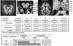
FDG-PET and 31P MRS studies in patients with intractable temporal lobe epilepsy (TLE) have found that the metabolic state of the thalamus is correlated with neuronal loss and gliosis in the ipsilateral hippocampus. The thalamic impairment is believed to result from recurrent seizures propagating through a network of involved structures. This hypothesis suggests that there should be a relationship between the metabolic state of the thalamus and ipsilateral hippocampus. In this study, we evaluated for this relationship using in vivo MRS. Spectroscopic images from n=20 controls and n=19 patients with TLE were acquired at 4T using a volume head coil. Voxels spanning each hippocampus (n=5) and thalamus (n=6 ) were reconstructed using anatomical landmarks to maximize consistency of sampling. In the thalamus, anterior locus 6 bilaterally is the most metabolically depressed. Additional significant declines were seen from ipsilateral loci 2-6. As expected there were statistically significant reductions in NAA/Cr (p[lt]0.05 to p[lt]0.001) in the ipsilateral and contralateral hippocampi in TLE patients in all loci except the most posterior location of the contralateral hippocampus (data not shown). To assess the inter-relationships between the hippocampus and thalamus in patients, we determined the mean hippocampal NAA/Cr (averaging loci 2-5) with the mean anterior thalamic NAA/Cr (averaging loci 4-6) (Table). The strongest correlation is between the ipsilateral hippocampus and the ipsilateral anterior thalamus. The distribution of neuronal injury/loss is greatest in the ipsilateral anterior thalamus, and is well correlated with injury in the ipsilateral hippocampus. This is consistent with the major hippocampal efferent (fornix) bilaterally projecting to the anterior thalamic nuclei, the dorsal medial and lateral dorsal nuclei. The extent of NAA/Cr decline in ipsilateral loci 4-5, and 2-3 is markedly less than that seen in locus 6, and may reflect the intra-limbic and intra-thalamic connectivity that may propagate neuronal injury. These data support the model that the impairment and damage associated with recurrent seizures as measured by NAA originating in the ipsilateral hippocampus propagates through the network of involved structures and results in corresponding impairment/damage in the contralateral hippocampus and bilateral thalami[figure1] (Supported by NIH PO1 NS39092, RO1 NS40550, Dana Foundation.)
