The Relationship Between Quantitative MRI and Seizure Outcomes After Temporal Lobe Epilepsy Surgery
Abstract number :
1.335
Submission category :
9. Surgery / 9A. Adult
Year :
2018
Submission ID :
502567
Source :
www.aesnet.org
Presentation date :
12/1/2018 6:00:00 PM
Published date :
Nov 5, 2018, 18:00 PM
Authors :
Marcia E. Morita-Sherman, Cleveland Clinic; Ruta Yardi, Cleveland Clinic; William Bingaman, Cleveland Clinic; Jorge A. Gonzalez-Martinez, Epilepsy Center, Neurological Institute, Cleveland Clinic; Imad M. Najm, Cleveland Clinic Epilepsy Center; Stephen Jo
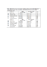
Rationale: The treatment of choice for drug-resistant epilepsy is brain surgery. MRI is a critical tool to localize the epileptic pathology. Recent data obtained using complex imaging research protocols have suggested that subtle atrophy in brain regions outside the surgical bed may also play an important role in seizure outcome. We explore here the prognostic value of volumetric MRI measurements in the context of temporal lobe surgery, using an FDA-approved software available for routine clinical practice. Methods: We hypothesize that subtle structural evidence of epileptic network pathology extending beyond the temporal lobe reduces the odds of seizure freedom after surgery. We identify quantitative MRI (qMRI) predictors of outcome defined by Engel I using NeuroQuant, an automatic qMRI measurement tool available in the clinical setting, providing percentile volume data of 138 brain regions against an age and gender-matched normative cohort. We retrospectively reviewed 99 patients (50 women) who had temporal lobe resection for the treatment of drug-resistant focal epilepsy in our center from 2010 to 2013. We collected Clinical variables through chart review. We obtained quantitative MRI measurements of the preoperative MRI from NeuroQuant. We analyzed the RIGHT and LEFT sided resections separately, aiming to identify qMRI predictors of surgical outcome. Results: The comparison between quantitative volumes of different brain regions of patients with Engel I (n=65) versus Engle II-IV (n=34) in both groups (left-sided (n=51) and right-sided surgery (n=48)) showed that in the right-sided surgery group contralateral limbic network involvement, contralateral hemispheric atrophy and extent of the atrophy beyond the typical border of the resection could be predictors of Engel II-IV surgical outcome. In the left-sided surgery group, the contralateral involvement of frontotemporal regions and ipsilateral hemispheric atrophy could be predictors of Engel II-IV seizure outcome (Table 1). Upon qualitative visual analysis of the pre-operative MRI in the same study cohort, the presence of a lesion correlated with better outcomes in the LEFT sided resection group (Table2). Conclusions: Our findings suggest that the presence of subtle brain atrophy in ipsilateral areas that extend beyond the typical surgical border of a temporal lobe resection, as well as contralateral involvement of frontotemporal regions (left-sided surgical group) and contralateral limbic and hemispheric involvement (right-sided surgical group), might predict a poor surgical outcome. Further efforts are needed to study the role of quantitative imaging and its incorporation in routine clinical practice in the context of epilepsy surgery outcome prediction. Funding: R01 NS097719-01; A Nomogram to Predict Seizure Outcomes after Resective Epilepsy Surgery, PI Jehi

.tmb-.png?Culture=en&sfvrsn=29008f85_0)